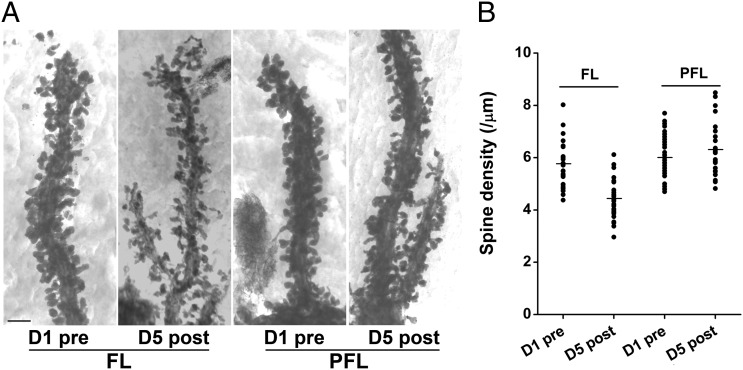
Fig. 4.
LTA is accompanied with elimination of PC spines in the FL. (A) Representative high-voltage EM images of PC spines along individual dendritic segments in the FL and PFL of control (D1 pre) and trained (D5 post) mice. (Scale bar, 2 μm.) (B) Pooled data showed selective spine elimination by 30% in the FL but not in PFL on day 5. Data were presented as mean ± SEM, **P < 0.01, vs. D1 pre, Student t test.

