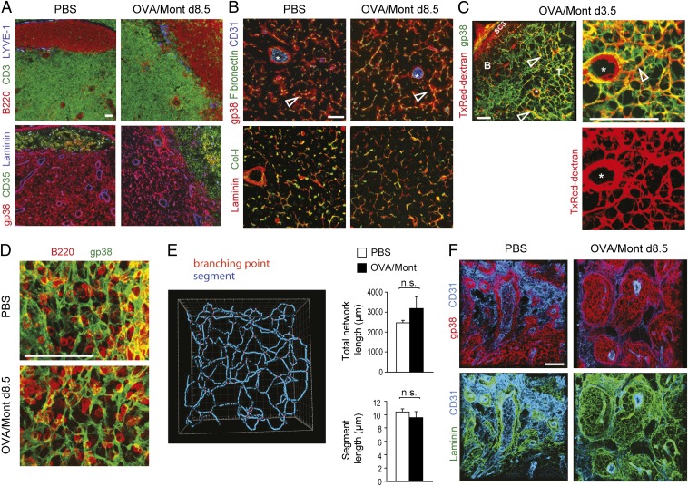
Fig. 4.
An expanding FRC network preserves its usual structure and function while extending into medullary cords. Immunofluorescence microscopy of cryostat sections (A and B) or 80-μm-thick vibratome sections (C–F) from draining LNs of PBS- or OVA/Mont-immunized mice were labeled with the indicated antibodies. (A) B220+ B cells and CD3+ T cells indicate B and T zones, respectively. LYVE-1 stains lymphatic vessels. Consecutive sections show FRC (gp38+CD35−) and FDC (gp38+/−CD35+) networks, as well as the laminin-positive basement membranes of vessels and conduits. (B) Higher-magnification image of the T zone showing CD31+ HEVs (asterisks) and reticular FRCs (gp38+) wrapped around fibronectin-positive conduits (open arrows). Conduits are composed of collagen I-positive (Col-I) fibrils surrounded by a laminin-positive basement membrane. (C) Texas (Tx) Red-dextran was injected s.c. at 3.5 d after OVA/Mont immunization, draining LNs isolated 30 min thereafter, followed by their processing for histological labeling. Open arrows highlight TxRed-dextran–positive conduits surrounded by gp38+ FRCs; the asterisk denotes an HEV with a perivascular space rich in TxRed-dextran. (D) Vibratome sections of the LN T zone in PBS- and OVA/Mont-immunized mice demonstrating a similar density and architecture as for the gp38+ FRC network. B220+ B cells are shown for a size comparison. (E) Filament tracer software was used to quantify the total network length and segment length of individual FRCs in images derived from gp38-labeled vibratome sections (mean ± SD), as shown in D. (F) Vibratome sections showing medullary cords displaying extensive gp38+ reticular FRC networks wrapping around laminin-positive structures and connecting with CD31high HEVs. The cords are demarcated by a thin layer of CD31intgp38+ lymphatic endothelium. Data are representative of two to three experiments with at least two mice and six LNs per mouse. (Scale bars: 100 μm.)

