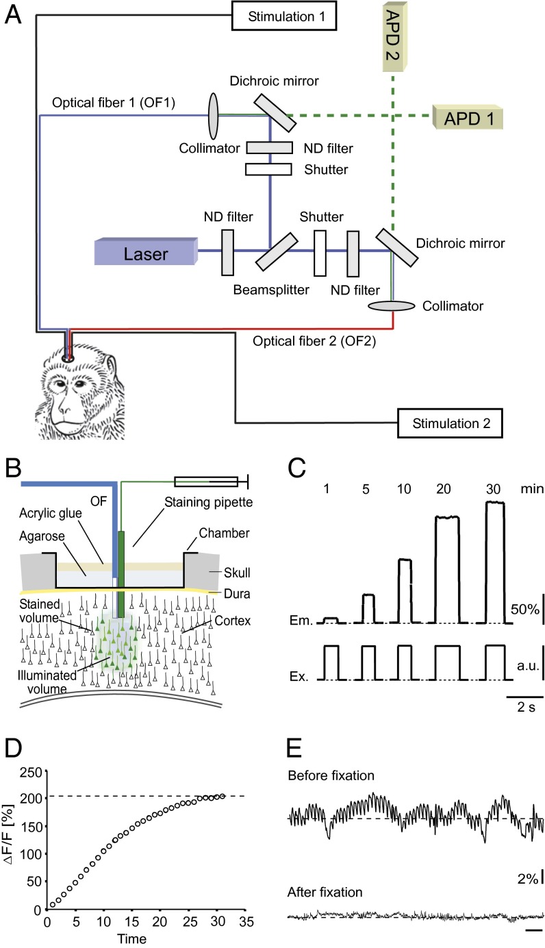
Fig. 1.
Experimental arrangement and calcium dye loading in monkey brain in vivo. (A) Scheme of the recording setup with two optical fibers combined with two microelectrodes for stimulation of the neurons and detection of spike activity. APD, avalanche photo diode; ND, neutral density. (B) Arrangement for multicell bolus loading of the tissue consisting of the staining pipette for application of the Ca2+-sensitive dye solution and an optical fiber. The green area indicates the region stained with the dye and the blue area, the volume of the tissue illuminated by the excitation light delivered from the optical fiber. (C) Fluorescence emission (Em) of the tissue after application of the dye containing solution monitored with the optical fiber. The excitation energy (Ex) given in arbitrary units (a.u.) was identical at all time points. (D) Plot of the increase of the fluorescence versus time after dye application. (E) Background fluorescence signal of the stained tissue before and after fixation of the optical fiber with agarose and acrylic glue.

