Figure 1.
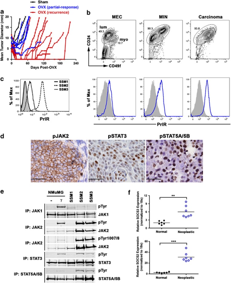
STAT1−/− mammary tumor cells exhibit persistent activation of JAK2, STAT3 and STAT5A/5B. (a) STAT1−/− mice bearing primary mammary tumors were sham-operated (black) or OVX (blue and red). Each line represents an individual tumor. (b) Disaggregated MECs and neoplastic cells from MIN (the earliest morphologically identifiable lesions) and carcinomas were gated based on CD49f and CD24 levels (top; circles). PrlR expression on CD49fint CD24hi luminal (lum) epithelial cells was quantified using b-Prl and SA-PE (bottom). Representative flow cytometry plots from three independent STAT1−/− mice are shown. (c) ERα+ SSM2 and SSM3 cells displayed elevated PrlR expression compared with the control ERα− SSM1 cells. (d) Phosphorylated JAK2, STAT3 and STAT5 were observed in primary STAT1−/− mammary tumor cells using immunohistochemical analyses. Representative images of five primary mammary tumors are shown. Scale bar=33 μm. (e) Immunoprecipitation western blotting analysis of JAK1, JAK2, STAT3 and STAT5 in NMuMG, SSM1, SSM2 and SSM3 cells. IFNγ-stimulated NMuMG was used as a positive control. (f) SOCS3 and SOCS2 mRNA levels were quantified in nontransformed STAT1−/− mammary glands (normal) and STAT1−/− mammary tumors (neoplastic). **P<0.001, ***P<0.0001

