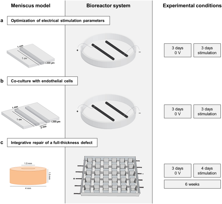
Figure 1. Electrical stimulation of meniscus.
(a) Optimization of electrical stimulation parameters in a micropatterned 3-D hydrogel system for cell migration. Inner or outer meniscus cells were encapsulated on plastic slides in a 1.8% fibrin channel (3.5 × 10⁶ cells/mL) and covered by a second layer of 1.8% fibrin to enable migration. After 3 days of pre-culture, slides were transferred into custom bioreactors with carbon electrodes spaced 2.5 cm apart, for 3 days of stimulation. (b) Co-culture of meniscus cells with endothelial cells. Human umbilical vein endothelial cells (HUVECs) and inner or outer meniscus cells (3.5 × 10⁶ cells/mL) were individually encapsulated on slides in parallel fibrin channels, left and right, respectively, and cultured for 3 days of pre-culture and 3 days of stimulation. (c) Juvenile bovine meniscus explants were punched with central cores of 1.5 mm diameter and immediately replaced to simulate a full-thickness defect. Explants were stimulated four days a week over six weeks of culture in a custom bioreactor system, consisting of a 5 × 6 array with carbon electrodes spaced 1 cm apart.

