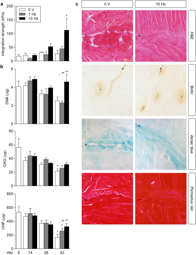
Figure 4. Electrical stimulation enhanced integrative repair and maintained the biochemical composition of explants.
(a) Integration strength of full-thickness defects in meniscus explants over six weeks with (3 V/cm, 1 Hz, 2 ms or 3 V/cm, 10 Hz, 0.2 ms pulse duration) and without stimulation (0 V). By day 42, stimulation at 10 Hz significantly promoted integrative repair versus explants without stimulation (0 V) and stimulated at 1 Hz. * p < 0.05 vs. day 0, vs. 0 V and 1 Hz at day 42; n = 3–9. (b) Biochemical composition of meniscus explants over six weeks: total DNA, sulfated GAG, hydroxyproline (OHP). At day 42, stimulation corresponded to significant upward trends in DNA and OHP content of explants compared to controls (0 V). Stimulation at 10 Hz maintained GAG and OHP content relative to day 0, in comparison to no stimulation (0 V) and stimulation at 1 Hz. * p < 0.05 vs. day 0; ** p < 0.04 for linear trend; n = 3–9. (c) Histological staining of meniscus explants at day 42: H&E for cell nuclei and cytoplasmic elements, BrdU for proliferating cells, Alcian blue for sulfated GAGs, Picrosirius red for collagens. The interface at the site of injury (arrows) appeared closely apposed with cells and newly synthesized matrix in explants stimulated at 10 Hz compared to no stimulation (0 V). Stimulated explants stained moderately more positive for BrdU+ nuclei, suggestive of enhanced cellular proliferation. Scale bar: 50 μm.

