Figure 3.
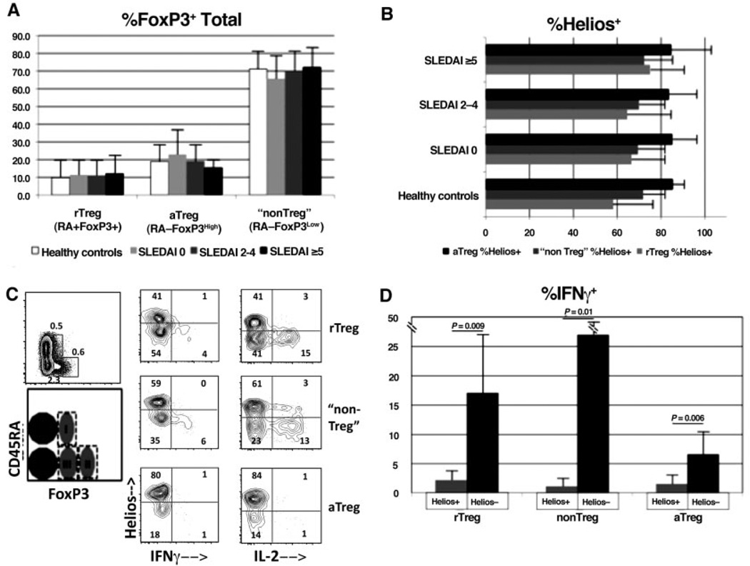
Presence of non-cytokine-producing FoxP3+Helios+ cells irrespective of the expression of CD45RA or the level of FoxP3 in healthy donors and patients with systemic lupus erythematosus (SLE). All panels are gated on CD4+ cells, and subtypes of FoxP3+ cells are based on the previously published system (18). A, Cell samples from 20 SLE patients and 17 healthy donors were stained for CD45RA in addition to CD4, Helios, and FoxP3, and the Treg cell subsets were determined as a percentage of the total CD4+FoxP3+ cells. rTreg = resting Treg cells; aTreg = activated Treg cells. B, Percentage of Helios expression in Treg cell subsets was determined in the samples shown in A. Patients in A and B were categorized according to Systemic Lupus Erythematosus Disease Activity Index [SLEDAI] scores. Values in A and B are the mean ± SD. C, Peripheral blood mononuclear cells from an SLE patient were directly stimulated ex vivo with 12-O-tetradecanoylphorbol-13-acetate (PMA)/ionomycin/GolgiStop, as described in Patients and Methods, prior to surface and intracellular staining. Costaining for the cytokines interferon-γ (IFNγ) and interleukin-2 (IL-2) was performed. Results are representative of cells from 6 different patients. Numbers in each compartment are the percentages of positive cells. Cell fractions I, II, and III are indicated. D, IFNγ production in 6 SLE samples was determined as described in C. Values are the mean ± SD. P values were determined by Student’s unpaired t-test.

