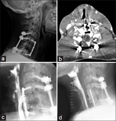
Figure 1.

Lateral cervical x-ray shows C4-T1 posterior fusion with C5-C7 ACDF and PEEK cage at the level of C6 with no apparent hardware defects (a) CT neck demonstrated air surrounding the implant at C6 as well as the anterior fusion plate (b) Barium swallow study showed a posterior esophageal barium leak at C5-C6 and a TE-fistula. The contents of the fistula are seen tracking inferiorly T1 and is seen anterior to the fusion plate (c) Barium swallow study done ten days postoperatively shows resolution of perforation (d)
