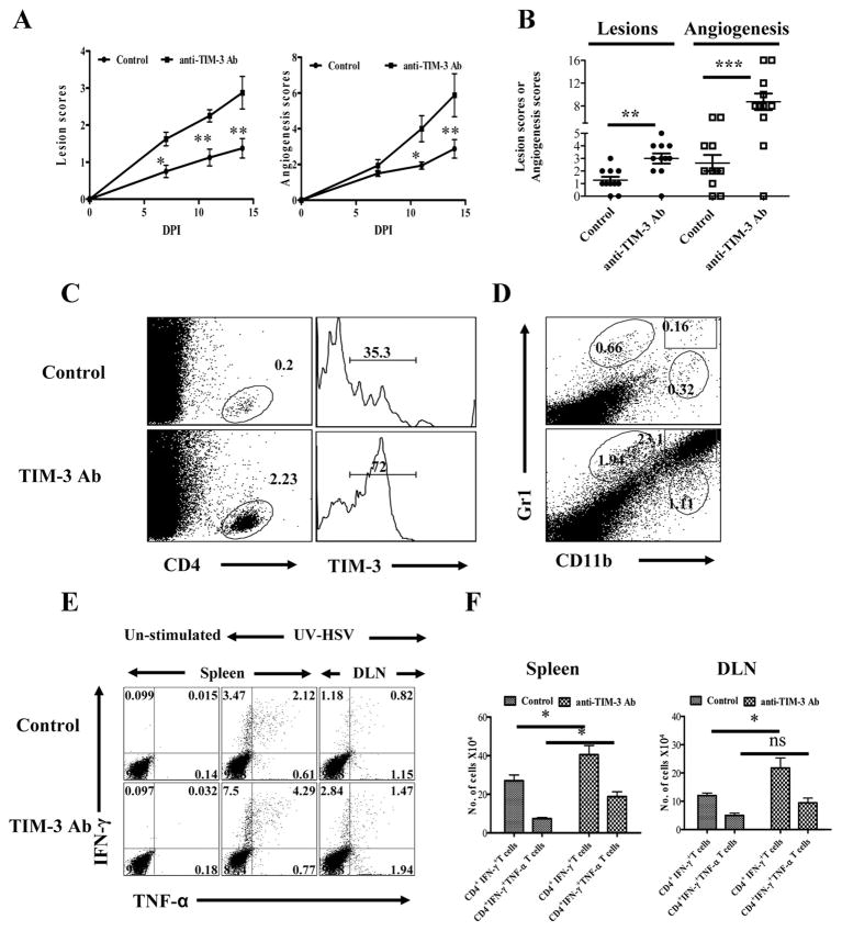
Fig. 2. Effects of TIM-3 blockade on corneal inflammation and HSV-specific CD4+ T cell immune response.
C57B/6 animals infected with 5×10⁵ PFU of HSV were given either anti-TIM-3 monoclonal antibody or isotype control antibody every alternate day starting from day 3 until day 13. The disease progression and immune parameters at day 14 were analyzed. A. The SK lesion severity and magnitude of angiogenesis is shown. B. Cumulative scores of lesion severity and angiogenesis at 14 days post infection. C. Percentages and phenotype (surface TIM-3) of CD4+ T cells in the corneas of control (upper panel) and antibody treated (lower panel) animals is shown D. CD11b+Gr1+ PMN infiltrated into cornea of control (upper panel) and antibody treated (lower panel) animals is shown. E. HSV-specific CD4+ T immune response in control and anti-TIM-3 antibody treated animals is shown at day 14. Dot plots depicting the percentages of IFN-γ+ TNF-α+ CD4+ T cells is shown. F. Total number of HSV-specific CD4+IFN-γ + and CD4+IFN-γ+TNF-α+ T cells in spleen and DLN at day 14 is shown.

