Fig. 4.
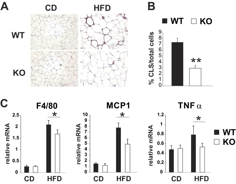
MLK3 deficiency reduces macrophage infiltration in visceral adipose tissue. A: representative images of hematoxylin and eosin-stained epididymal fat sections from WT and MLK3-KO mice. B: no. of crown-like structures (CLS) in epididymal fat sections from HFD-fed mice was calculated as %total cells. More than 1,000 cells/section were counted. Data are means ± SD; n = 8 mice. C: expression of F4/80, MCP-1, and TNFα was measured by quantitative RT-PCR analysis normalized to the expression of cyclophilin. Data are means ± SE; n = 8. **P < 0.01; *P < 0.05.

