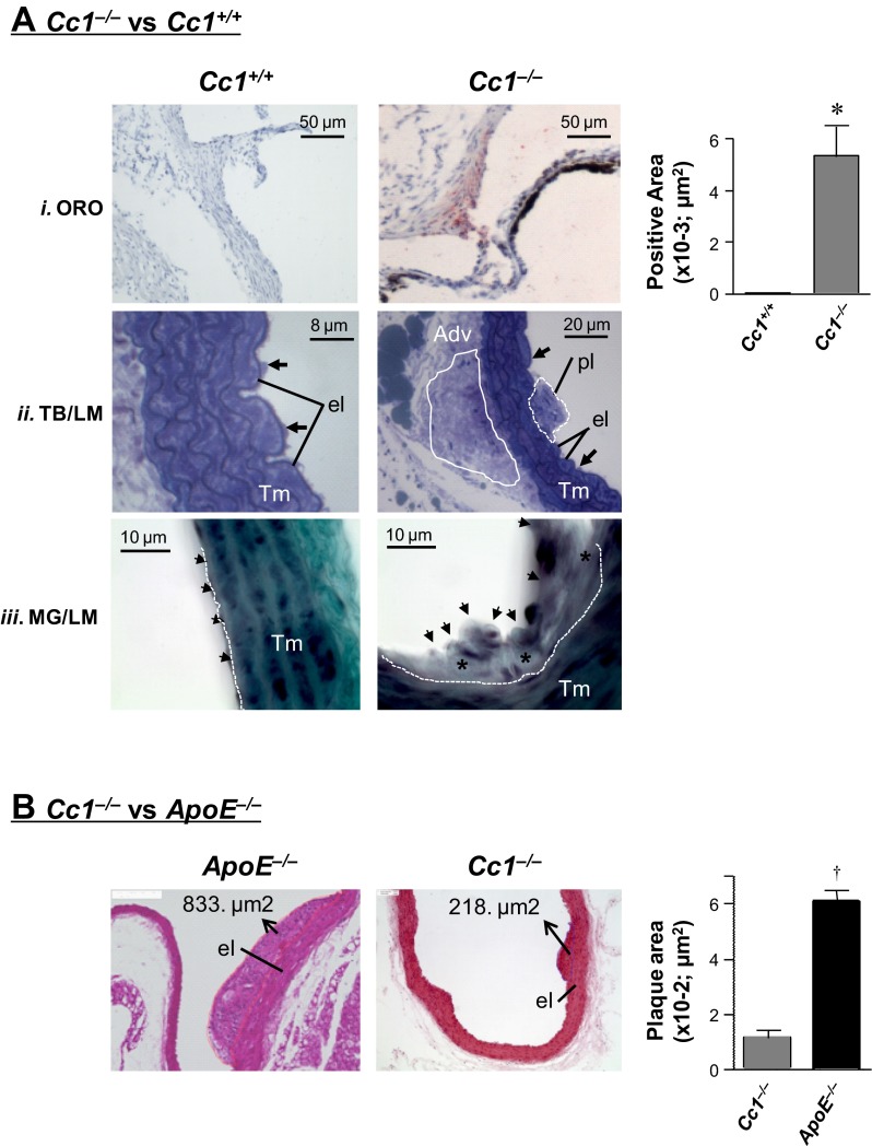
Fig. 1.
Morphological analysis of aortic lesions. A: staining of aortae from 6-mo-old male mice (n > 5 per group). A.i: cross-sections of aortic root stained with Oil red O (ORO) depicted lipid deposition in the intima of carcinoembryonic antigen-related cell adhesion molecule 1 (Ceacam1)-null (Cc1−/−) but not Cc1+/+ mice. Quantification of stained areas is represented in the side graph, *P < 0.05 vs. Cc1+/+. A.ii: light microscopic (LM) evaluation of Toluidine blue (TB)-stained semithin sections from aortic arch and ascending aorta revealed plaques in aortic intima of Cc1−/− aorta (dotted white line, pl) with increased cell density of aortic adventitia (solid white line, Adv) at the opposite site. Black arrows, Endothelial lining cells; Tm, tunica media. A.iii: LM evaluation of aortic arch sections stained by Masson-Goldner (MG) technique revealed a small atherosclerotic lesion with increased subendothelial deposition of fibrotic materials (*) in Cc1−/− aorta. Arrows, endothelial lining of aorta; white dotted line, border between the aortic intima and Tm. B: histological analyses of aortae from 8-mo-old mice (n = 3 per group), sectioned and H&E stained for plaque area measurement in 5 sections per mouse. el, Internal elastic membrane layer. Values are presented as means ± SE in ApoE−/− relative to Cc1−/− (†P < 0.01).

