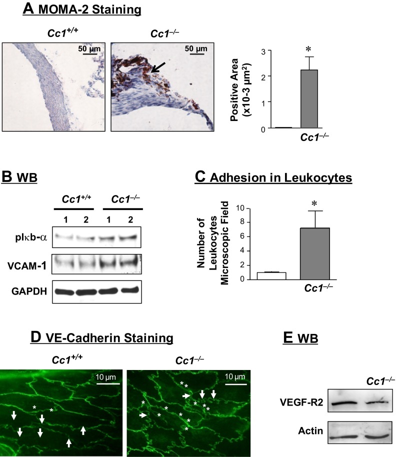
Fig. 2.
Vascular inflammation and leukocyte adhesion. A: immunostaining cross-sections of aortic root with monocyte and macrophage antibody 2 (MOMA-2; n > 5 of 6-mo-old mice per group). Stained area was measured and presented in the accompanying graph. Values are presented as means ± SE. *P < 0.05 vs. Cc1+/+. B: Western blot (WB) analysis of phospho-Iκbα and vascular cell adhesion molecule-1 (VCAM-1) content in aortic lysates from 6-mo-old mice, normalized against GAPDH. Gel represents 4 mice per group, but only 2 mice were included. C: aortic segments from 8-mo-old mice (n > 5) were incubated with isolated leukocytes for in vitro measurement of leukocyte adhesion. Values are presented as means ± SE. *P < 0.05 vs. Cc1+/+. D: en face VE-cadherin analysis of aortae (n > 5 of 6-mo-old mice per group). E: WB analysis of vascular endothelial growth factor receptor 2 (VEGFR-2) content in aortic lysates derived from 6-mo-old mice. Gel represents 3 mice per group.

