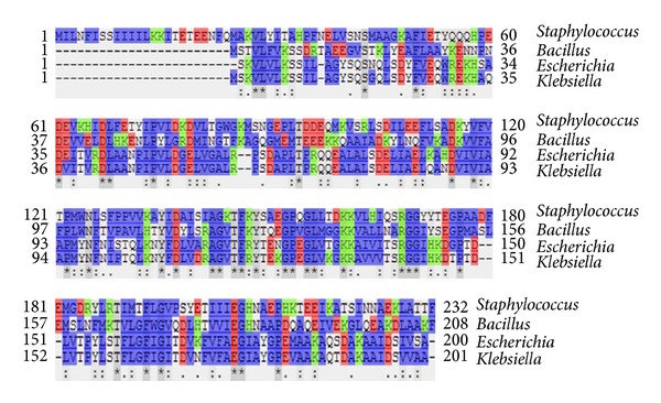
Figure 2.

Multiple sequence alignment of azoreductase from Staphylococcus aureus, Bacillus subtilis, Escherichia coli, and Klebsiella sp. Amino acid residues are colored to indicate their similarity: blue = hydrophobic, red = acidic, and green = basic residues. For comparative model, the sequence identity with the template is of 34%.
