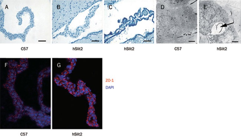
Figure 3.
Changes in the structure and the function of the choroid plexus. (A, B and C) The structure of the choroid plexus of a C57 mouse (A), a hSlit2 mouse (B) and a hSlit2 mouse with megahead (C). hSlit2 mice had structurally different choroid plexuses compared with C57 mice. Scale bar: 150 μm. (D and E) Electron micrograph of choroid plexus epithelial cells from the mouse lateral ventricle. Some vacuoles appear between the junctional complexes linking the epithelial cells in hSlit2 mice. Scale bar: 1.5 μm. (F and G) Immunofluorescence of the choroids plexus probed with an anti ZO-1 antibody. Cell-cell adhesion was different in hSlit2 mice compared with C57 mice.

