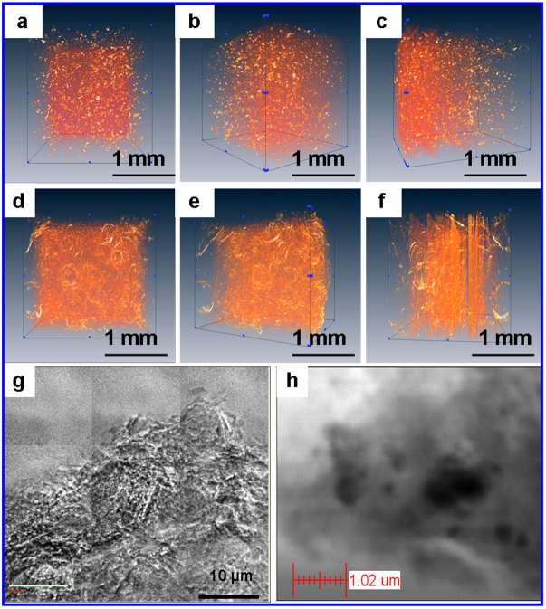
Figure 2. Microstructure of LCU by synchrotron radiation X-ray imaging system.
(a–c), Aggregate structure and distribution of natural attapulgite from three different angles by synchrotron radiation hard X-ray imaging system (SRHX). (d–f), 3D micro/nano network structure and distribution of ATP within LCU from three different angles by SRHX. (g,h), Micro/nano network structure of ATP (g) and Mg distribution (h) within LCU by scanning and transmission X-ray (soft) microscopy (STXM) on synchrotron radiation (1314 eV). The brighter regions in (g) correspond to ATP rods, and the organics (mainly urea and few P) are embedded within the ATP meshes.

