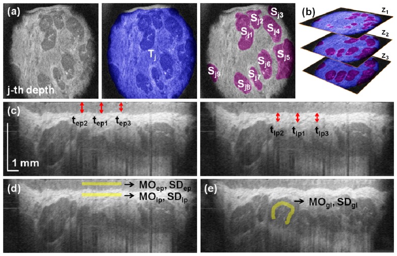
Fig. 2.
Quantitative assessment of the mucosa of the lower lip and LMSGs. (a) Observable mucosal tissue (blue) and individual glands (violet) were segmented to determine the corresponding areas. (b) The analyses were performed at three depths to provide with volumetric analysis. (c) Measurement of the average thickness of the epithelium (left) and the hyperreflective part of the lamina propria (right). (d) Estimation of the reflectivity and heterogeneity of the epithelium and the lamina propria. (e) Estimation of the reflectivity and heterogeneity of the glandular tissue.

