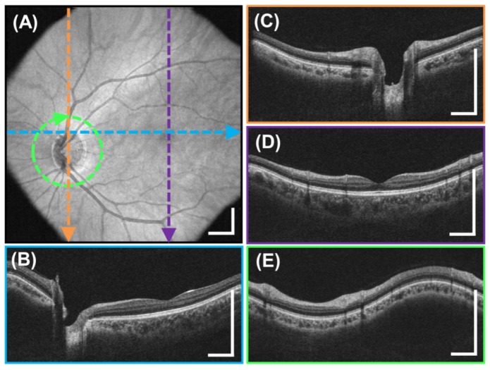
Fig. 8.
Motion-corrected, wide field 10 x 10 mm2, 350 x 350 A-scan volume generated from two raster scanned volumes acquired in 1.4 seconds each. (A) En-face OCT fundus image. (B-D) Color-indicated cross sections on the fundus. (E) Interpolated 3.4 mm diameter circumpapillary scan extracted from motion-corrected volumetric data. Scale bars are 1 mm.

