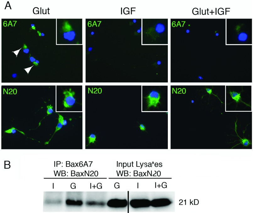
Figure 2. Detection of active Bax in OPCs exposed to glutamate.
Late OPCs were treated with glutamate (500 μM)±IGF-I (20 ng/ml) for 24 h. (A) Immunostaining for conformation-specific Bax (6A7; upper panels) or total Bax (N20; lower panels). Bax was detected using an AF488-conjugated secondary antibody (green); DAPI-stained nuclei are shown as blue. (B) Active Bax was detected by immunoprecipitation from glutamate-treated OPCs using conformation-specific Bax antibody (6A7) followed by Western blotting with total Bax N20 antibody (21 kDa). I, IGF-I; G, glutamate; I+G, IGF-I plus glutamate. Data are representative of two independent experiments at 24 h. Similar results were obtained at 18 h. Line in gel in (B) indicates that a blank lane was removed from the gel for publication purposes.

