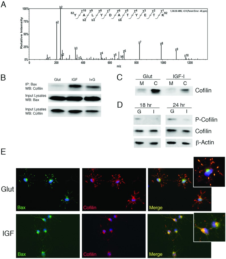
Figure 5. Identification of cofilin as a Bax-binding partner in IGF-I-treated OPCs.
(A) Bax was immunoprecipitated from OPCs treated for 24 h with IGF-I. The protein band corresponding to ~20 kDa was excised from the gel and subjected to ESI–LC–MS/MS analysis. Scaffold-generated MS/MS spectrum of a doubly charged ion corresponds to Y82ALYDATYETK92 peptide from cofilin-2 (NCBI Reference Sequence NP_001102452.2). The observed y- and b-ion series confirmed the peptide sequence. (B) Isolated protein from OPCs treated for 24 h with glutamate or IGF-I was used to immunoprecipitate total Bax (N20). Cofilin was detected by Western immunoblotting in IGF-I-treated OPCs. Association of cofilin with Bax was barely detectable in OPCs treated with glutamate at 24 h. Data are representative of two experiments; similar results were obtained at 18 h. (C) Analysis of cofilin in mitochondrial (M) and cytoplasmic (C) fractions 24 h following exposure of OPCs to glutamate or IGF-I. (D) Detection of p-cofilin (Ser9) in OPCs treated with glutamate (G) but not in OPCs treated with IGF-I (I) at 18 h and 24 h. (E) Immunodetection of Bax (N20; AF488-green) and cofilin (AF546-red) in OPCs treated with either glutamate (Glut) or IGF-I (IGF). DAPI (blue) was used to detect nuclei. Data are representative of at least two independent experiments.

