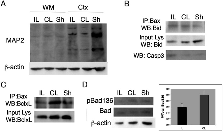
Figure 6. Bcl-2 family regulation in immature white matter after H–I.
H–I was produced in P6 Wistar rat pups by cauterizing the common carotid artery followed by systemic hypoxia for 75 min. Subcortical white matter (WM) was dissected at 24 h recovery from ipsilateral (IL), contralateral (CL) and Sham (Sh) operated brains and used for protein isolation (n=6 pooled samples). (A) Western blot of MAP-2 expression in cortex vs WM dissected regions. (B) Active caspase 3 increased in IL white matter (bottom panel). (B-C) Isolated protein was used to immunoprecipitate total Bax (N20). Bax-associated Bid (B) and Bcl-xL (C) were detected by Western immunoblotting. The data for the Bax–BclxL association are representative of three independent experiments and the data for the Bax–Bid association are from two independent experiments. (D) Detection of pBad(Ser136) and total Bad by Western immunoblotting of protein from IL, CL and Sh white matter. Histogram shows ratio of p-Bad/total Bad in IL vs CL white matter. Values represent averages±S.D. from three independent experiments; P = 0.08).

