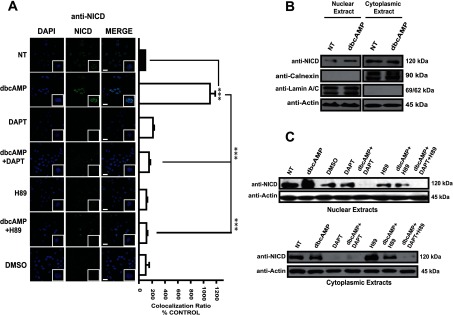
Figure 2. Only cleaved NICD is translocated from the cytoplasm to nuclei in a PKA- and γ-secretase-dependent manner.

In all cases, C6 cells were treated with dbcAMP (750 μM); where indicated, the γ-secretase inhibitor DAPT (40 μM) or the PKA inhibitor (H89) was added 30 min before dbcAMP treatment; 24 h after treatment, cells were processed. (A) Immunostaining for cleaved-NICD (Val1744; green); nuclei were counterstained with DAPI (blue). Images and co-localization ratios were obtained as in Figure 1 (scale bar=25 μm). (B and C) Subcellular fractionation and Western blot analysis using 75 μg of cytoplasmic or nuclear protein and anti-cleaved-NICD, anti-actin, anti-lamin A/C and anti-calnexin (as controls of nuclear and cytoplasmic extracts). Molecular masses are depicted on the right side. Representative images from at least three independent experiments are shown.
