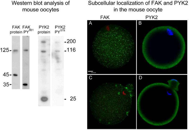
Figure 2. Detection and sub-cellular distribution of PYK2 and FAK kinases.

Western blot analysis of MII oocytes (left) was performed on groups of 60 oocytes/ lane probed with antibodies to FAK protein (FAK protein), antibodies to phosphorylated FAK (FAK PY861), anti PYK2 protein (PYK2 protein), or anti phosphorylated PYK2 (PYK2 PY579) as described in’ Materials and Methods’. The apparent molecular weight of each major band was calculated by comparison with molecular weight standards and is presented in the margins (arrows).
Sub-cellular localization of FAK and PYK2 protein (right) was performed on oocytes collected prior to fertilization in vitro (A, C) or during anaphase/ telophase II (B, D) then labeled with anti-PYK2 protein and anti-FAK protein antibodies not targeted to phosphorylation sites in order to establish the sub-cellular distribution of the entire pool of these kinases in the oocyte. Bound antibodies were detected with alexa 488-anti-rabbit IgG (green). Chromatin was stained with ethidium homodimer (red) or Hoechst 33258 (blue). Magnification is indicated by the bar which represents 10 μm.
