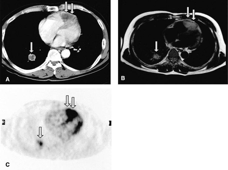
Figure 2.
Contrast-enhanced CT (A) and T1-weighted MRI (B) show a large intracardiac mass involving the pericardium and myocardium in the right ventricle (double arrows), and a mass in the right lower lobe of the lung (single arrows). 18F-FDG PET (C) demonstrates that the intracardiac tumor had an intensely increased FDG uptake (double arrows), which is strongly suggestive of malignancy.

