Abstract
The combination of idiopathic thrombocytopenic purpura (ITP) and chronic renal failure (CRF) is uncommon. This report highlights a case of renal transplantation in a patient with ITP. A 35-year-old man with ITP was admitted with uremic symptoms. A renal transplant and splenectomy was simultaneously performed. A prophylactic pneumococcous vaccination was performed and intravenous immunoglobulin (1 g/kg) was administered before and after the operation. The patient's platelet count increased gradually after the splenectomy. During a two-year follow up period, the graft function was well maintained. Renal transplantation in a patient with ITP is recommended with a well-designed strategy to prevent potential complications.
Keywords: Idiopathic thrombocytopenic purpura, Renal transplantation
INTRODUCTION
Idiopathic thrombocytopenic purpura (ITP) is an autoimmune disorder characterized by a low platelet count and mucocutaneous bleeding. Patients with this ailment are at risk of spontaneous bleeding, and the major cause of fatal bleeding is intracranial hemorrhage1).
The combination of ITP and chronic renal failure (CRF) is uncommon, and renal allograft transplantation in a patient having these manifestations is especially rare. Only one case has been reported in Japan, in 19862).
This report covers our experience performing renal transplantation in a patient with ITP.
CASE REPORT
A 35-year-old man was hospitalized for generalized weakness and exertional dyspnea. He had developed ITP at 15 years of age and chronic glomerulonephritis at 27 years of age. However, he had been well without receiving a definite treatment.
Upon admission, his vital signs were stable except for an increased blood pressure (150/100 mmHg), and a physical examination was unremarkable. His laboratory data were as follows: hematocrit, 24.4% (normal range, 42% to 52%); hemoglobin, 8.1 g/dL (normal range, 13 to 18 g/dL); white blood count, 6,100/µL (normal range, 4,000 to 10,500/µL); platelet count, 12,000/µL; serum blood urea nitrogen, 86.9 mg/dL (31.0 mmol/L) (normal range, 9 to 20 mg/dL [3.2 to 7.1 mmol/L]); and serum creatinine, 7.71 mg/dL (681.6 µmol/L) (normal range, 0.8 to 1.5 mg/dL [71 to 133 µmol/L]). His 24-hour urine protein excretion was 7.6 g. Blood chemistry showed a serum sodium level of 142 mmol/L (normal range, 135 to 145 mmol/L); potassium, 4.6 mmol/L (normal range, 3.5 to 5.5 mmol/L); albumin, 3.4 g/dL (normal range, 3.8 to 5.0 g/dL); calcium, 7.9 mg/dL (normal range, 8.3 to 10 mg/dL); and phosphate 4.6 mg/dL (1.49 mmol/L) (normal range, 2.5 to 5.0 mg/dL [0.80 to 1.60 mmol/L]). Liver function tests were all within normal ranges. All serologies were negative or normal, including antinuclear antibodies, anti-neutrophil cytoplasmic antibodies, hepatitis B surface antigen and hepatitis C antibody. Serum immunoglobulin levels were normal and C3 and C4 complement levels were normal.
Urinalysis revealed a specific gravity of 1.010, pH 7.5, 3+ protein, and macrohematuria. A chest X-ray was negative and a renal ultrasound revealed normal-sized kidneys. Bone marrow biopsy findings were compatible with ITP.
The patient was initially treated with steroids (1 mg/kg/day) for two weeks, but there was no increase in his platelet counts. Meanwhile, the blood urea nitrogen level increased gradually (to 55.3 mmol/L), and his uremic symptoms (nausea, poor oral intake and generalized weakness) became aggravated. Hemodialysis was initiated, and renal transplantation and a splenectomy were scheduled to be performed simultaneously as the optimal management of end stage renal disease and ITP.
The donor was the patient's 28-year-old sister, and HLA typing confirmed a haploidentical donor-recipient match. Before renal transplantation, a prophylactic pneumococcal vaccination was performed and intravenous immunoglobulin (IVIG, 1 g/kg) was given one day before transplantation. Cyclosporine, prednisolone, and mycophenolate mofetil (MMF) were used as initial immunosuppressive agents. At the time of renal transplantation, his platelet count was 89,000/µL.
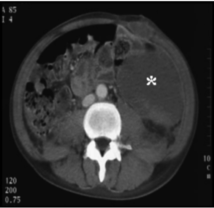
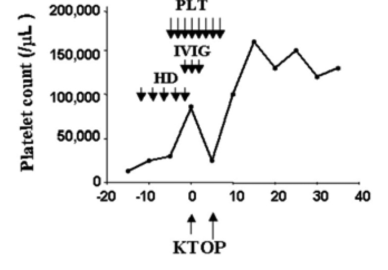
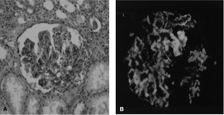
The patient's initial graft function was good. Intraabdominal bleeding had developed just after transplantation, but bleeding was not observed thereafter (Figure 1). His platelet count gradually increased on the tenth post-transplant day and the patient was discharged on the 37th post-transplant day with a normal serum creatinine level (114 µmol/L) and a platelet count of 124,000/µL (Figure 2). The native kidney biopsy findings were consistent with IgA nephropathy (Figure 3).
Figure 1.
Abdominal computed tomography image. Note the huge hematoma (asterisk) in the abdominal cavity.
Figure 2.
Clinical course. Note the gradual increase in platelet count after renal transplantation. Key: PLT, platelet concentrates; IVIG, Intravenous immunoglobulin; HD, hemodialysis; KT, kidney transplantation; OP, Operation for hematoma removal.
Figure 3.
Renal biopsy findings. (A) This photomicrograph shows widening of the mesangial area and mesangial cells (H & E stain, original magnification, ×400). (B) Immunofluorescent microscopy shows a deposit of immunoglbulin A on the mesangial area.
Two months later, the patient was readmitted because of fever, chill and headache. Cryptococcus neoformans was recovered from the blood, cerebrospinal fluid and pleural fluid. Systemic cryptococcosis was well-controlled with the reduction of immunusuppressants and an intravenous amphotericin B treatment.
A two-year follow-up of the patient showed a favorable clinical course. His platelet count was stable and the graft function was well maintained. No further overwhelming sepsis was observed.
DISCUSSION
The current report highlights a successful renal transplantation in a patient with ITP and CRF. The ideal outcome for this case would have been an initial positive response to the steroid treatment, a successful renal transplant without bleeding, and a stable platelet count with immunosuppression after renal transplantation. However, this patient showed a poor response to steroids, and experienced bleeding and systemic cryptococcosis after renal transplantation. Based on this experience, we conclude that transplantation is possible in a patient with ITP and CRF, but that a well-designed strategy is needed to prevent possible complications.
The first decision made was to choose the best treatment for CRF (dialysis versus transplantation) in this patient because of his uremic symptoms due to renal failure (blood urea nitrogen 86.9 mg/dL and creatinine 7.71 mg/dL). In determining the treatment, our major concern was how to decrease the bleeding complications. Dialysis treatment (hemodialysis and peritoneal dialysis) is known to partially improve hemostatic defects in uremia, but a complete recovery is unexpected3). Therefore, we recommended renal transplantation to reduce the risk of possible fatal bleeding.
Our next consideration was how to treat ITP in this uremic patient while he waited for a renal transplantation. In general, the treatment of ITP consists of steroid and cytotoxic drug administration and a spelectomy. Steroid treatment is the first line of treatment but it has a low and late response and a high relapse4). Compared to steroid therapy, a splenectomy shows an early (one week) and positive response rate, and sustained effects. On the other hand, intraoperative bleeding and postoperative sepsis are unfavorable complications. In this case, steroid therapy was given as an initial treatment for two weeks, but there was no response. Moreover, it aggravated the uremic symptoms by increasing blood urea nitrogen levels, and there was concern that excessive immunosuppression before transplantation might increase the later risk of infection. We therefore, stopped steroid therapy and decided to perform a splenectomy.
The timing of the splenectomy and the prevention of intraoperative bleeding were other important factors while treating this patient. Initially, sequential operations (renal transplantation after a splenectomy) were scheduled because a simultaneous operation might have increased his risk of bleeding, and the expected recovery of a platelet count after a splenectomy would have provided safer conditions for the transplant. However, performing a splenectomy in a patient with uremic condition also carries a high risk of bleeding, and we were unable to predict whether this patient would respond satisfactorily5). Therefore, we decided on simultaneous operations to avoid the risk of repeated bleeding.
IntraVenous ImmuneGlobulin (IVIG) therapy is recommended to decrease surgical risks in patients with ITP, and a dose of IVIG (1 g/kg) before platelet transfusions usually brings the platelet count up rapidly6). Indeed, Takahara et al. reported a successful renal transplantation with immunoglobulin therapy in a patient with ITP2). In our patient, IVIG (1 g/kg) was administered one day before renal transplantation with two more subsequent doses, but his platelet count did not recover immediately. Consequently, he experienced intraoperative bleeding and needed an operation for hematotma removal. A retrospective review showed that the patient's platelet count gradually increased, suggesting a positive response to the splenectomy. Based on this experience, we recommend sequential operations (a splenectomy before renal transplantation) to reduce the risk of bleeding.
A splenectomy predisposes individuals to risks of overwhelming infections5). These are most often due to encapsulated organisms, especially pneumococci, Hemophilus influenzae type b, and meningococci, but any similar agents may cause fulminant infections7). In general, pneumococcos, H. influenzae type b and meningococcal vaccines are recommended at least two weeks before a splenectomy. Our patient had received a pneumococcal vaccination before his splenectomy. He showed no immediate infections, but suffered from systemic cryptococcosis two months later. In this case, it is difficult to define the association between the splenectomy and the cryptococcal infection, but excessive immunosuppression using MMF coupled with the splenectomy might have been responsible for the fungal infection7).
The cause of the chronic renal failure in this case is interesting. The biopsy obtained from the operation revealed IgA nephropathy. Until now, the association between IgA nephropathy and ITP has not been defined clearly and only three reports have been published that cover this matter8-10). Two cases showed simultaneous IgA nephropathy and ITP, and one case showed the development of IgA nephropathy after a splenectomy. Therefore, it is not clear whether ITP is responsible for the development of IgA nephropathy. In this patient, ITP developed in his childhood and IgA nephropathy developed later. It is therefore possible that impaired IgA clearing, due to splenic dysfunction, is responsible for IgA nephropathy.
A two-year follow-up of this case has shown a favorable clinical course. Platelet counts were maintained within normal ranges, and there was no consequential episode of acute rejection or overwhelming sepsis. The former may be related to the splenectomy and subsequent immunosuppression with cyclosporine and the steroid treatment, and the latter may be related to the well-matched HLA transplant and to the splenectomy. Therefore, the clinical outcome for a patient with IPT may be good if the complications occurring during the early post-transplant period can be overcome.
In summary, we report a successful renal transplantation in a patient with CRF and ITP. If a splenectomy is planned, sequential rather than simultaneous operations seem to be a better approach, and minimal immunesuppression is recommended to avoid overwhelming sepsis. Our experience may thus, be helpful to manage patients with ITP waiting for renal transplantation.
References
- 1.Frederiksen H, Schmidt K. The incidence of idiopathic thrombocytopenic purpura in adults increases with age. Blood. 1999;94:909–913. [PubMed] [Google Scholar]
- 2.Takahara S, Ichikawa Y, Ishibashi M, Takaha M, Sonoda T. Renal transplantation and idiopathic thrombocytopenic purpura. Clin Transpl. 1986:133. [PubMed] [Google Scholar]
- 3.Weigert AL, Schafer AI. Uremic bleeding: pathogenesis and therapy. Am J Med Sci. 1998;316:94–104. doi: 10.1097/00000441-199808000-00005. [DOI] [PubMed] [Google Scholar]
- 4.Lechner K. Management of adult immune thrombocytopenia. Rev Clin Exp Hematol. 2001;5:222–235. doi: 10.1046/j.1468-0734.2001.00043.x. [DOI] [PubMed] [Google Scholar]
- 5.Cines DB, Blanchette VS. Immune thrombocytopenic purpura. N Engl J Med. 2002;346:995–1008. doi: 10.1056/NEJMra010501. [DOI] [PubMed] [Google Scholar]
- 6.Bussel JB, Pham LC. Intravenous treatment with gammaglobulin in adults with immune thrombocytopenic purpura: review of the literature. Vox Sang. 1987;52:206–211. doi: 10.1111/j.1423-0410.1987.tb03029.x. [DOI] [PubMed] [Google Scholar]
- 7.Bisharat N, Omari H, Lavi I, Raz R. Risk of infection and death among post-splenectomy patients. J Infect. 2001;43:182–186. doi: 10.1053/jinf.2001.0904. [DOI] [PubMed] [Google Scholar]
- 8.Watanabe M, Fujimoto T, Iwano M, Shiiki H, Nakamura S. Report of a patient of primary Sjogren syndrome, IgA nephropathy and chronic idiopathic thrombocytopenic purpura. Nihon Rinsho Meneki Gakkai Kaishi. 2002;25:191–198. doi: 10.2177/jsci.25.191. [DOI] [PubMed] [Google Scholar]
- 9.Ikeda K, Maruyama Y, Yokoyama M, Kato N, Yamanoto H, Kaguchi Y, Nakayama M, Shimada T, Tojo K, Kawamur T, Hosoya T. Association of Graves' disease with Evans' syndrome in a patient with IgA nephropathy. Intern Med. 2001;40:1004–1010. doi: 10.2169/internalmedicine.40.1004. [DOI] [PubMed] [Google Scholar]
- 10.Morino M, Inami K, Shibuya A, Sasaki N. IgA nephropathy and idiopathic thrombocytopenic purpura with splenectomy: a case report. Pediatr Nephrol. 1994;8:345–346. doi: 10.1007/BF00866358. [DOI] [PubMed] [Google Scholar]


