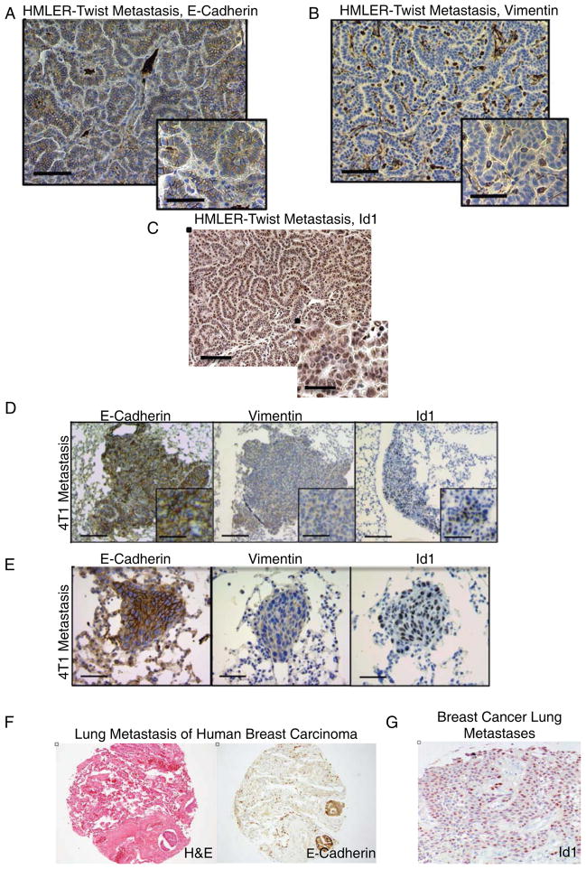
Figure 2. Id1 Expression is Associated with an Epithelial Phenotype in Breast Cancer Lung Metastases.
(A) IHC of lung metastases from HMLER-Twist cells injected mice reveals tumor cells positive for E-Cadherin and negative for Vimentin (B). Scale bar = 200μm, inset scale bar = 100μm.
(C) Id1 staining in HMLER-Twist lung metastases. Scale bar = 200μm, inset scale bar = 100μm.
(D) 4T1 cells form lung metastases positive for Id1 and E-Cadherin, and negative for Vimentin as determined by IHC. Scale bar = 500μm, inset scale bar = 100μm
(E) IHC for Id1, E-Cadherin, and Vimentin in macrometastases 5 days after 105 4T1 cells were injected into the tail vein of Balb/c mice. Scale bar = 200μm.
(F) Hematoxylin-eosin and E-Cadherin staining of human lung parenchyma showing two tumor emboli of metastatic breast carcinoma.
(G) IHC for Id1 in lung metastasis of human breast arcinoma.

