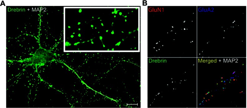
Figure 1. Drebrin, GluN1 and GluA2 clusters as markers for spines and spine subtypes.
(A) Representative image of DIV14 neuron showing drebrin clusters as spine markers. MAP2 was used as a dendrite marker. (B) Representative quadruple-stained dendrite segment showing debrin clusters co-localized with or without GluN1 or GluA2 clusters. In merged image: drebrin (green), GluN1 (red), GluA2 (blue), and MAP2 (white). Scale bar 10 μm.

