
Figure 3.
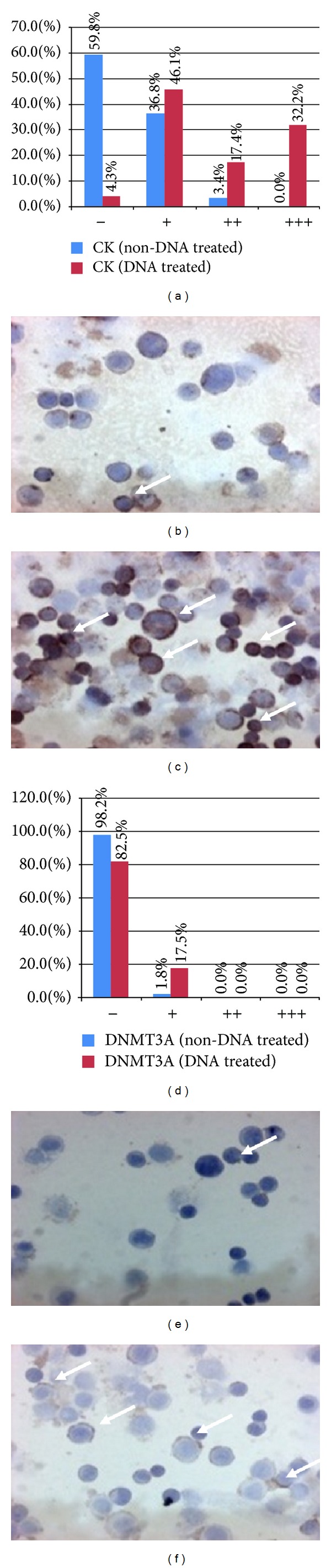
Distribution of cytokeratine (CK; (a)–(c)) and DNA methyltransferase 3A (DNMT3A; (d)–(f)) immunoreactive HT29 cells. (a) and (d) x-axes represent the intensities of immunoreactions from negative (−) to strong (+++). y-axes represent the distribution (percentage) of cells showing different immunoreaction intensities (blue columns: control cells; red columns: type 3. DNA-treated cells). (b) and (c) CK expression before (b) and after (c) type 3. DNA incubation (immunoreactive cells/arrows/display brownish cytoplasmic reaction, 200x magnification, hematoxylin costaining). (e) and (f) DNMT3A expression before (e) and after (f) type 3. DNA incubation (immunoreactive cells/arrows/displayed brownish cytoplasmic reaction, 200x magnification, hematoxylin costaining).