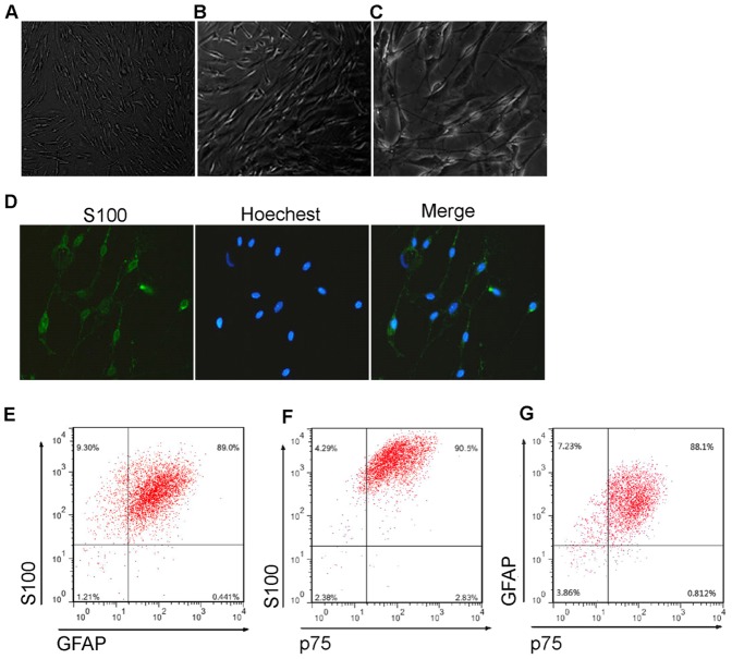
Figure 2. Isolation and Characterisation of mouse SCs to obtain the conditioned medium.
(A) Phase-contrast micrographs of SCs isolated from mouse sciatic nerve and dorsal root ganglia. The cells had typical spindle-shaped SC morphology. (B) Immunofluorescence staining demonstrated the isolated cells expressed SC marker S100 protein. (C) Flow cytometry assay showed that most of the cells were S100 (96.77±1.46%), GFAP (92.92±4.94%), and p75 (93.38±0.90%) positive, and 86.12±1.53% of the cells expressed all of the three SC markers (data are mean % cells±S.E.M.).

