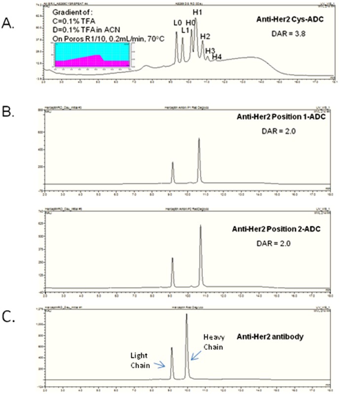
Figure 2. Antibody HPLC analysis.
The reversed phase HPLC of deglycosylated reduced and denatured (A) Anti-Her2 Cys-ADC, (B) Anti-Her2 Position 1-ADC, Anti-Her2 Position 2-ADC and (C) Anti-Her2. Insert shows the gradient profile and chromatography conditions. There were two drugs/antibody determined by the intact MS analysis or one drug per heavy chain. The reversed phase HPLC profile of the cysteine conjugated anti-Her2 ADC shows the number of drugs conjugated to the heavy (H) and light (L) chains determined by the intact mass spectrometry analysis.

