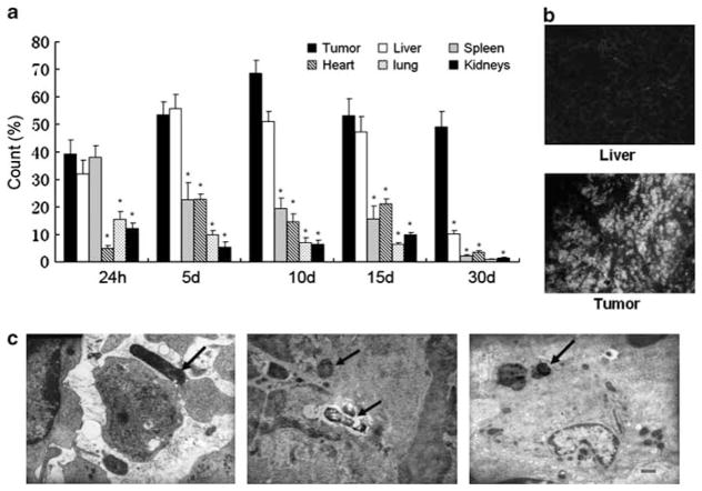
Figure 1.
Recombinant S. Typhimurium distribution in C57/BL6 tumor-bearing mice. (a) By using GFP expression as a marker, the bacteria distribution in different organ tissues at specified times after inoculation of bacteria (*P<0.01). (b) The GFP expression was observed in tumor and liver tissues by fluorescence microscope. (c) Electron microscopic analyses of S. Typhimurium infecting H22 tumors. Left panel: bacteria in the intercellular substance of tumor tissues, middle panel: bacteria in the cellular nucleus that was being autolyzed, right panel: bacteria in the tumor-associated macrophage. The arrows indicate the bacteria.

