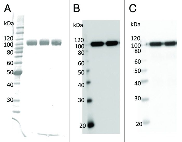
Figure 2. Electrophoretic mobility and immunoblot analysis of purified pp-PA83. (A) Coomassie-stained SDS-PAGE. pp-PA83 was loaded at 1 µg/lane. (B and C) Immunoblot. pp-PA83 was loaded at 100 ng/lane. pp-PA83 was separated by SDS-PAGE, transferred to polyvinylidene fluoride membranes and blotted with (B) a commercially available anti-4 × His mAb or (C) a pp-mAbPA.
