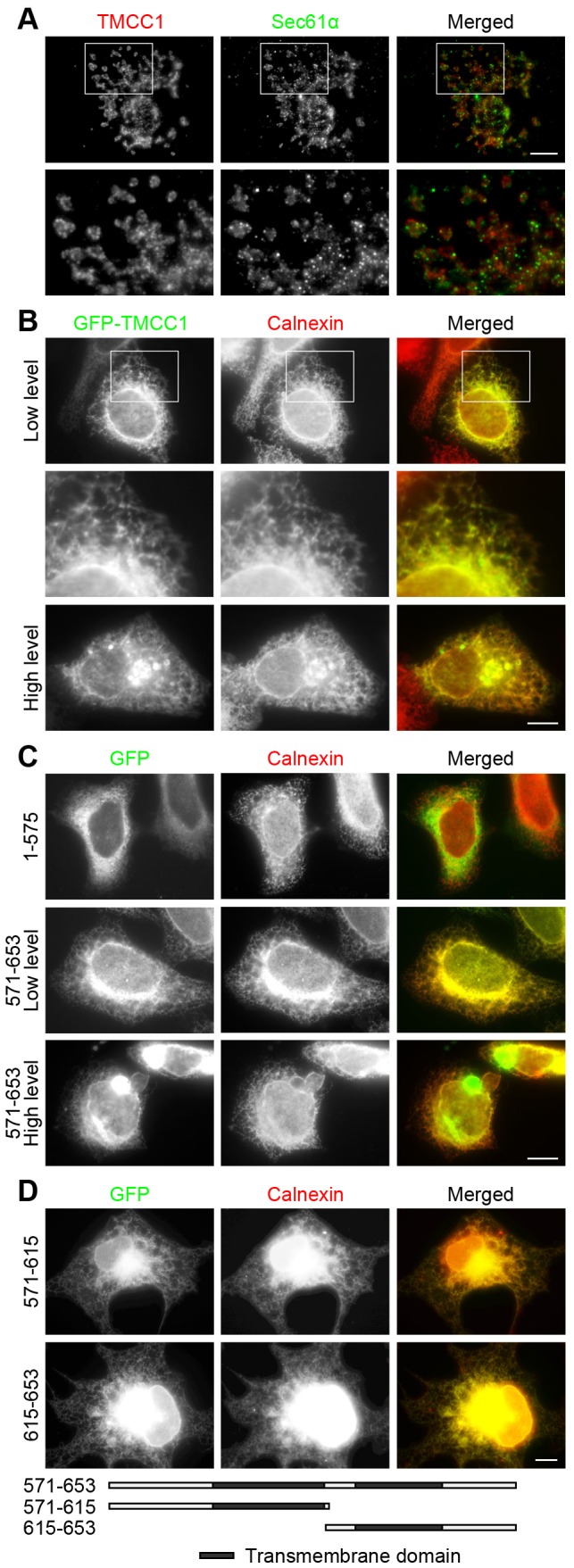
Figure 3. Subcellular localization of TMCC1.

(A) Saponin-extracted COS-7 cells were fixed with methanol and stained with both Sec61α and TMCC1 antibodies; the boxed area shown is magnified. (B–C) HeLa cells were transfected with plasmids encoding GFP-tagged TMCC1 full-length protein, TMCC1(1–575), or TMCC1(571–653); 24 h post-transfection, cells with low and high levels of the exogenous proteins were fixed with methanol and stained with an anti-calnexin antibody. A magnified view of the boxed area in (B) is shown. (D) COS-7 cells were transfected with plasmids encoding GFP-tagged TMCC1(571–615) or TMCC1(615–653); 24 h post-transfection, cells were fixed with paraformaldehyde and then permeabilized with 0.2% Triton X-100 for 10 min at room temperature. Cells were then stained with an anti-calnexin antibody. Scale bars, 10 µm.
