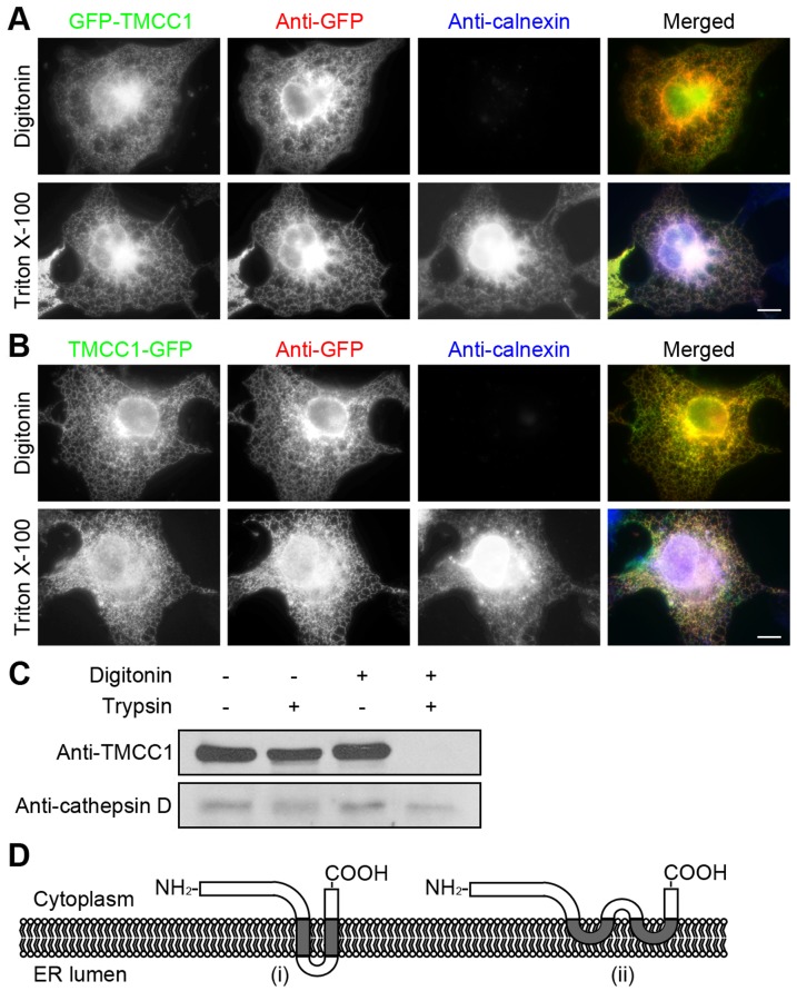
Figure 5. Topology of TMCC1.
(A–B) COS-7 cells were transfected with a plasmid encoding N-terminal (A) or C-terminal (B) GFP-tagged TMCC1; 24 h post-transfection, cells were fixed with paraformaldehyde and then permeabilized with either 40 µg/mL digitonin for 5 min on ice or 0.2% Triton X-100 for 10 min at room temperature. Cells were then co-stained with GFP and calnexin antibodies. Scale bars, 10 µm. (C) HeLa cells were treated with various combinations of digitonin and trypsin and then immunoblotted for TMCC1 and cathepsin D. (D) Two possible models of TMCC1 topology. Model (i) shows a transmembrane topology with 2 transmembrane domains, and Model (ii) shows an intramembrane topology with 2 intramembrane domains.

