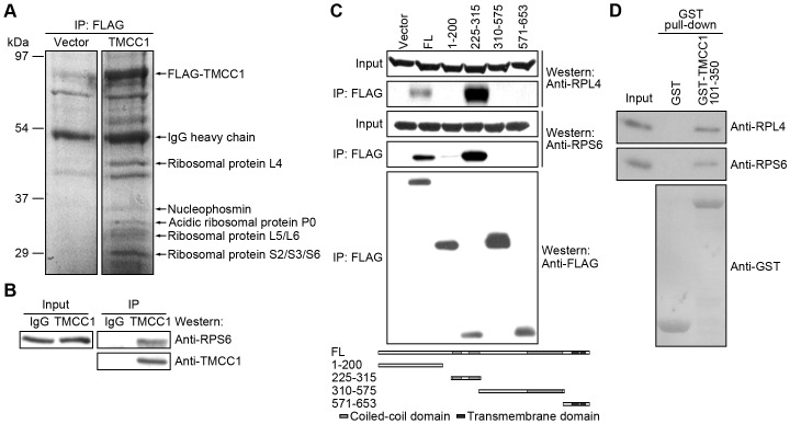
Figure 7. Interaction of TMCC1 with ribosomal proteins.
(A) HEK293T cells were transfected with FLAG-tagged TMCC1 plasmid or the vector; 24 h post-transfection, cell lysates were prepared for anti-FLAG immunoprecipitation. Immunoprecipitated proteins were visualized on the protein gel by staining with Coomassie Brilliant Blue R-250. The protein bands marked in the figure were identified by mass spectrometry. Vector, pFLAG-CMV2 vector. (B) HeLa cell lysates were collected for TMCC1 immunoprecipitation, and samples were immunoblotted for TMCC1 and the ribosomal protein RPS6. (C) HEK293T cells were transfected with plasmids encoding FLAG-tagged TMCC1 full-length protein or fragments; 24 h post-transfection, cell lysates were collected for anti-FLAG immunoprecipitation. Ribosomal and FLAG-tagged proteins were analyzed by western blotting. Vector, pFLAG-CMV2 vector. FL, full-length TMCC1. (D) Ribosomes prepared from HeLa cells were incubated with purified GST or GST-TMCC1(101–350) protein and then pulled down using GSH-beads; ribosomal and GST-tagged proteins were analyzed by western blotting.

