
Figure 4. Comparison of atrial contents of collagen I and collagen III in atrial tissues from AF and SR subjects.
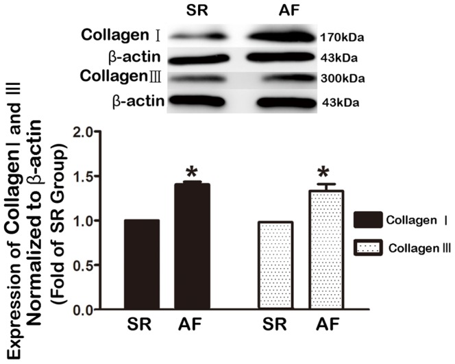
Collagen contents were determined by Western blot analysis. Left panel: Typical examples of Western blot bands showing upregulation of both collagen I and collagen III; right panel: averaged band densities, normalized to β-actin. Note that the collagen contents were substantially higher in atrial tissues from AF patients than from SR controls. SR, sinus rhythm; AF, atrial fibrillation. *P<0.05; n = 10.