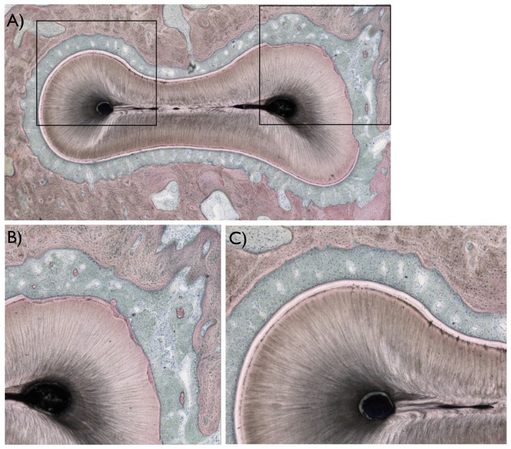
Figure 4. Histologic images of the areas of interest observed in the study.
(a) A descriptive histologic section showing the region of interest evaluated (marked in black). (b) For all teeth evaluated, damage to the root was observed primarily at the buccal and furcation region of the roots, where regions of cementum were removed in full thickness and root dentin removed in partial thickness. (c) In regions not surgically affected, the typical anatomic features of hard (alveolar bone, cementum, and root dentin), and soft tissues (periodontal ligament) were observed.

