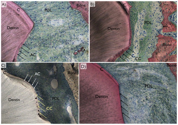
Figure 7. Representative histologic images depicting the periodontal ligament (PDL) region between bone and the tooth, where original cementum thickness (C) could be depicted in the vicinity of a defect.
The three different regeneration patterns comprised (a) acellular cementum (AC), (b) cellular cementum (CC), (c) mixed cellular and acellular cementum (MCA), or (d) no cementum regeneration (arrows).

