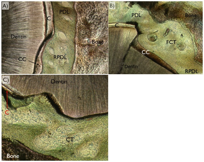
Figure 8. For periodontal ligament regeneration assessment, optical microscopy in circular polarized mode was utilized and the different tissues (dentin, bone, cementum [C], cellular cementum [CC], acellular cementum [AC], original periodontal ligament [PDL], regenerated periodontal ligament [RPDL], and fibrous connective tissue [FCT were easily depicted).
Among the three variations of healing pattern observed were (a) full regeneration, (b) partial regeneration, and (c) no regeneration (no fibrous bridging between bone and cementum along the defect perimeter) were observed.

