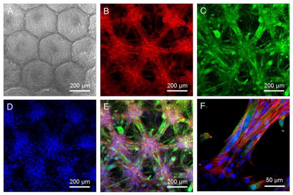
Figure 5.
C2C12 myoblasts cultured in a PLGA SHAIP in a differentiation medium for 10 days. A) A transmission bright-field optical micrograph showing the cells grown in the scaffold. B–D) CLSM images showing (B) f-actin, (C) myogenin, which is a marker for differentiated myoblasts, and (D) nuclei. E) Superimposed image of (B–D). F) An enlarged view of the superimposed image showing multinucleated myotubes connecting the cell bodies in adjacent pores.

