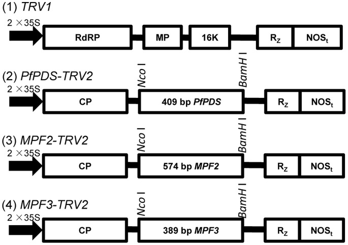
Figure 1. Organization of TRV-VIGS vectors.
TRV cDNA clones were placed in between duplicated CaMV 35S promoters (2×35 S) and the nopaline synthase terminator (NOSt) in a T-DNA vector [22]. RdRp: RNA-dependant RNA polymerase; MP: movement protein; 16 K: cysteine rich protein; Rz: self-cleaving ribozyme; CP: coat protein. PfPDS-, MPF2-, and MPF3-specific fragments were inserted separately into TRV2 using Nco I and BamH I restriction sites.

