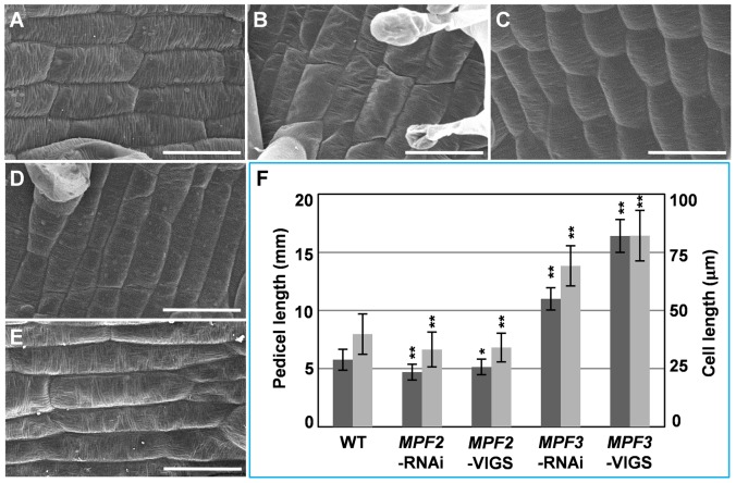
Figure 5. MPF3 and MPF2 regulate pedicel development and pedicel cell length.
Pedicel cells from the WT (A), MPF2-RNAi (B), MPF2-VIGS (C), MPF3-RNAi (D), and MPF3-VIGS (E). Bars = 50 µm. (F) Quantification of the pedicel size (dark gray column) and the respective pedicel cells lengths (light gray column). The number of pedicels analyzed was 20 for each line above. The numbers of cells analyzed were 60 in WT, MPF2-RNAi and MPF2-VIGS, and 50 in MPF3-RNAi and MPF3-VIGS. Mean values and standard deviation are presented in both cases. Two-tailed t-test significance was given as follows: one star for p<0.05, and two stars for p<0.01.

