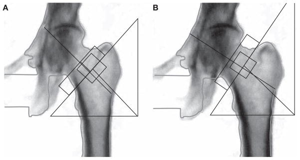
Figure 3.

Femoral neck box placement. Each manufacturer has its own standards for correct femoral neck box placement. The dual-energy X-ray absorptiometry technologist must be familiar with the recommendations for the instrument that is used and place the neck box in the same position in serial studies. Here, both images are the same, with the femoral neck box correctly placed on the right. If these were different scans done to evaluate possible changes over time, any comparison would be invalid. (A) Incorrect analysis: femoral neck T-score = −3.2. (B) Correct analysis: femoral neck T-score = −3.0.
