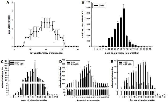
FIGURE 4.
Myelin Ag is first detected in microglia in the CNS during EAE. C57BL/6 mice were immunized with MOG35-55 peptide for induction of EAE as previously described. Animals were monitored and disease scored daily. (A) Representative mice were scored for EAE and sacrificed as indicated over the course of EAE (n = 3 per day) and brain tissue frozen and sectioned. Tissue sections were stained as indicated and evaluated by confocal microscopy analysis for (B) CD4+ T cells, (C) CD11c+, (D) CD11b+, or (E) Ly-6G+ cells. Cells in (C–E) were also stained with anti-MBP mAb. Numbers shown represent mean ± S.D., ANOVA (p≤0.002).

