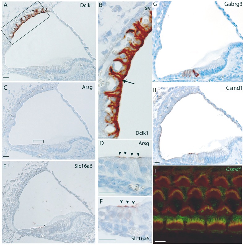
Figure 2. Immunohistochemistry in the mouse cochlea at P5 (P4 for Slc16a6 gene).
Brown indicates positive staining. A, B) Expression of Dclk1, showing intense staining in the marginal cells including projections towards the basal cells of the stria vascularis (arrows in A,B); C, D) Expression of Arsg is localized at the top of sensory hair cells in the organ of Corti (bracket in C, arrowheads in D). E, F), Apical hair cells at P4, showing staining in outer hair cells of Slc16a6 of the organ of Corti (bracket in E, arrowheads in F). Note that these samples are from the C3HeB/FeJ strain, which is pigmented. Gabrg3 shows a striking specific expression in the outer and inner hair cells (arrowheads in H). In particular, the inner hair cells have the strongest staining. H, I) Expression of Csmd1 is localized at the top of sensory hair cells in the organ of Corti (arrowheads in H; I) confocal expression shows a strong staining localized in the stereocilia of inner hair cells and weak staining is also present in the stereocilia of outer hair cells. Csmd1 is labelled in green while rhodamine/phalloidin labels actin filaments of stereocilia in red. Merged image (yellow) indicates colocalization of Csmd1 with actin in stereocilia hair bundles. Scale bars: A–H: 20 µm; I: 5 µm.

