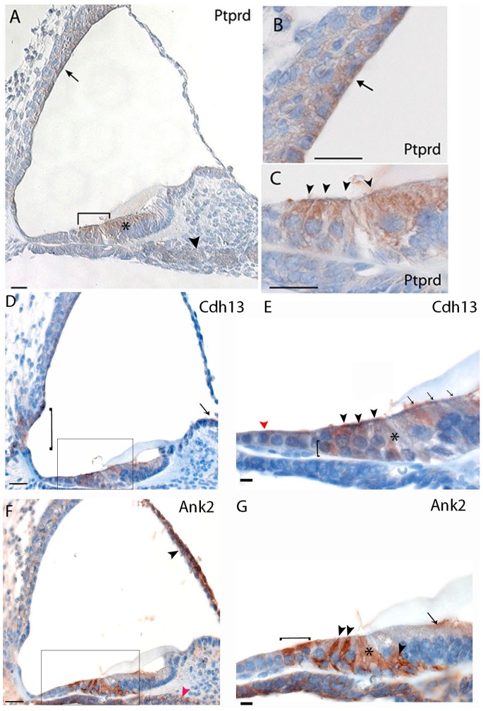
Figure 3. Immunohistochemistry in the mouse cochlea at P5.
Brown indicates positive staining. A, B, C) Ptprd is localized in hair cells of the organ of Corti (bracket in A, arrowheads in C), in the marginal cells of the stria vascularis (arrow in A, B), in the supporting cells of the Kölliker’s organ (marked by an asterisk in A) and in the spiral ganglions neurons (arrowhead in A); D, E) Cdh13 is expressed in cells of Claudius (red arrowhead in E), outer and inner hair cells (arrowheads in E), Deiters’ cells (bracket in E) and pillar cells (asterisk in E), cells of the Kolliker’s organ (arrows in E) in the organ of Corti. Staining was also noted in interdental cells (arrow in D), stria vascularis (asterisk in D), spiral prominence and external sulcus cells (bracket in D). F, G) Ank2 could be noted in the Hensen’s cells (bracket in G), Deiters’ cells (arrowheads in G)and pillar cells (asterisk in G) in the organ of Corti. Moreover, Ank2 is expressed in the Reissner’s membrane (arrowhead in F) and cells of the Kolliker’s organ (arrow in G). Scale bars: A–C: 20 µm. D, F: 20 µm; E,G: 10 µm. Rectangles label the regions shown in higher magnification.

