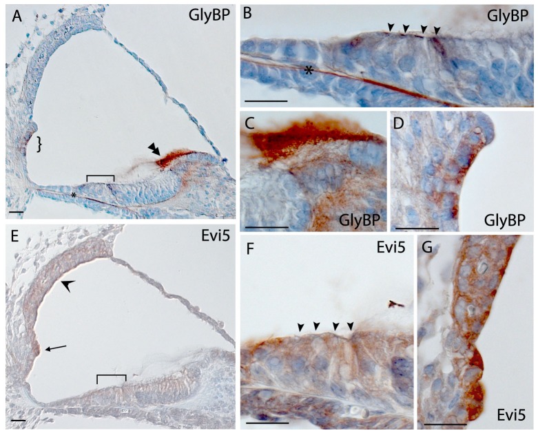
Figure 5. Immunohistochemistry in the mouse cochlea at P5.
Brown indicates positive staining. A, B, C, D) Expression of GlyBP showing staining in hair cells (bracket in A, arrowheads in B), tectorial membrane (double arrowhead in A, C), root cells (curly brackets in A, D) and basilar membrane (asterisk in A and B). E, F, G) Staining of Evi5 localized throughout the cochlea, including hair cells (bracket in E, arrowheads in F) and spiral prominence and stria vascularis (arrow and open arrowhead in A respectively, G). Scale bar indicates 20 µm.

