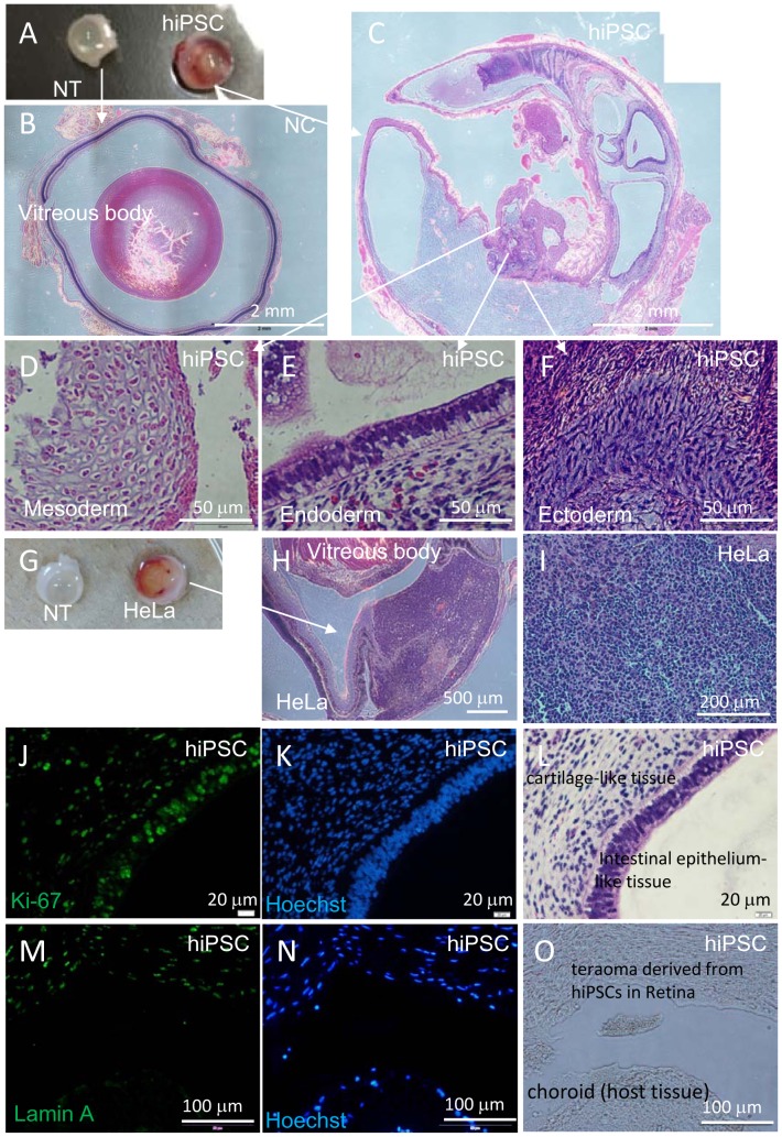
Figure 6. Histological analyses of hiPSCs or HeLa cells transplanted into the subretinal space of nude rats.
Eye balls were excised from a nude rat 7 weeks after subretinal transplantation of hiPSC. Non-transplanted right eye ball (NT) and left eye ball transplanted with 1×10⁴ hiPSCs (hiPSC) (A). HE staining of cross section of NT eye ball (B) and hiPSC-transplanted eye ball (C). HE staining of hiPSC-derived teratoma with three germ layers: cartilage-like tissue (mesoderm, D), intestinal epithelium-like tissue (endoderm, E) and neuron-like tissue (ectoderm, F) in hiPSC-transplanted eye ball. (G – O) Eye balls were excised from a nude rat 5 weeks after subretinal transplantation of HeLa cells. Non-transplanted right eye ball (NT) and left eye ball transplanted with 1×10⁵ HeLa cells (HeLa) (G). HE staining of cross section of HeLa cell-transplanted eye ball (H) and HeLa-derived tumor tissue (I). Anti-Ki-67-antibody (J), Hoechst 33258 (K) and HE staining (L) of serial sections of hiPSC-derived teratoma. Anti-Lamin A antibody (M), Hoechst 33258 (N) staining and microscopic image (O) of serial cross sections containing a boundary of hiPSC-derived teratoma and host rat tissue. Anti-Lamin A antibody specifically recognizes human cells in rat tissue.

