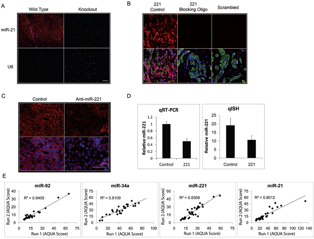
Figure 2. miRNA qISH assay validation.
(A) miR-21 and U6 ISH (red) on miR-21 knockout mouse heart tissue or wild type mouse heart tissue merged with DAPI (blue). (B) Representative examples of miR-221(red) and Scrambled probe (red) ISH performed on TMAs with and without the miR-221 blocking oligo merged with DAPI (blue) and Cytokeratin (green). (C) miR-221 ISH (red) performed on MCF-7 cells transfected with 30 nM control (anti-miR negative control) or anti-miR-221 inhibitor and merged with DAPI (blue). (D) Quantification of miR-221 knockdown in MCF-7 cells by qRT-PCR normalized by U6 and qISH from 24 random fields normalized by scrambled probe. (E) Reproducibility of miR-92a, miR-34a, miR-221, and miR-21 qISH performed on different days (run 1 versus run 2) on near serial sections of the breast cancer index TMA. Scale bars represent 50µm.

