Figure 4.
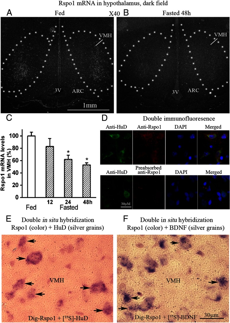
Dark field photomicrograph of hypothalamus, double in situ hybridization, and immunofluorescence. A, Fed rat. B, Fasted 48-hour rat. 3V, third brain ventricle; C, Densitomitry analysis of Rspo1 mRNA levels in VMH of fed and fasted rats. *, P < .05 compared with fed rats; n = 5. D, Photomicrograph from VMH, green represents HuD protein, red represents Rspo1 protein, blue represents nuclei. Yellow shows colocalization. E, Color represents Rspo1 mRNA, silver grains represent HuD mRNA. Arrow heads indicate colocalization. F, Purple color represents Rspo mRNA, silver grains represent BDNF mRNA n = 5. DAPI, 4',6-diamidino-2-phenylindole, dihydrochloride.

