Figure 6.

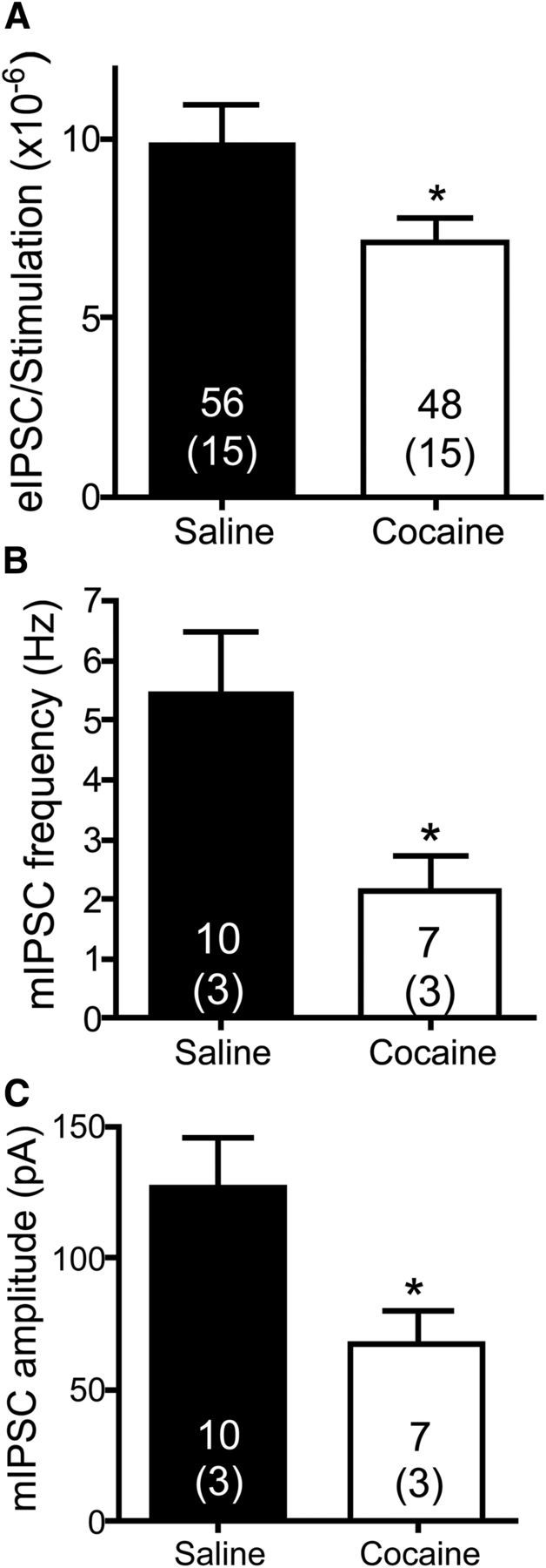
GABA neurotransmission in the VP is depressed in cocaine-extinguished rats. A, GABA IPSC amplitudes (pA) were divided by the stimulation amplitude (μA) used to generate them. The eIPSC/stimulation ratio was higher in yoked saline rats [9.87 ± 1.1 (×10−6)] than in cocaine-extinguished rats [7.10 ± 0.69 (×10−6)], indicating that for a given stimulation amplitude the resultant eIPSC was lower in cocaine-extinguished rats (unpaired two-tailed t test, t(102) = 2.059, p = 0.042). Number of cells presented above number of rats (in parentheses). B, The frequency of mIPSCs was significantly lower in cocaine-extinguished rats (2.15 ± 0.6 Hz) compared with yoked saline rats (5.43 ± 1 Hz), indicating a tonic presynaptic inhibition of GABA neurotransmission (unpaired two tailed t test, t(15) = 2.97, p = 0.010). C, The amplitude of mIPSCs was significantly lower in cocaine-extinguished rats (67.8 ± 12.2 pA) compared with yoked saline rats (127.0 ± 18.8 pA), indicating postsynaptic inhibition of GABAergic receptors (unpaired two-tailed t test, t(15) = 2.77, p = 0.014). *p < 0.05 compared with yoked saline.
